
Psychotherapy changes the brain by driving neuroplasticity: it reshapes functional connectivity and synaptic strength in emotion-regulation circuits, producing durable symptom improvement that shows up on brain scans. Meta-analyses of neural mechanisms in depression and anxiety consistently report post-therapy decreases in anterior cingulate cortex, inferior frontal gyrus, and insula activation, alongside measurable improvements in emotional regulation. These are not metaphorical changes. They are physical, observable shifts in how your brain processes emotion, threat, and self-referential thought, which is why psychotherapy is increasingly understood as a biological intervention, not merely a conversational one.
- Therapy drives neuroplasticity by strengthening adaptive neural pathways through repeated, emotionally engaged practice.
- Neuroimaging studies show partial normalization of depression and anxiety-related brain patterns after successful treatment.
- Psychotherapy's relapse-reduction effects often outlast those of medication alone, pointing to durable structural and functional reorganization.
- The brain regions most consistently affected include the prefrontal cortex, amygdala, anterior cingulate cortex, insula, and hippocampus.
Why neuroplasticity is the foundation of how therapy changes you
Neuroplasticity refers to the brain's capacity to reorganize itself by forming new synaptic connections, remodeling dendritic spines, and shifting patterns of functional connectivity between regions. It is not a single event but a continuous process, one that experience drives at every stage of life. When you learn something new, practice a skill, or process a difficult emotion in a safe relational context, your brain physically changes in response.
The Nobel Prize-winning neuroscientist Eric Kandel was among the first to articulate why this matters for psychotherapy. His core argument: learning modifies synaptic connections and alters circuit function, and psychotherapy is, at its root, a structured learning process. As he and colleagues put it:
"Insofar as psychotherapy works by producing stable changes in behavior, it presumably does so through learning, by producing changes in gene expression that alter the strength of synaptic connections and structural changes that alter the anatomical pattern of interconnections between nerve cells of the brain."
— Eric Kandel, as cited in Rebuilding the brain with psychotherapy
This framing dissolves the old distinction between "psychological" and "biological" treatments. Every mental process is a brain process. When a therapist helps you reframe a fear-based belief or process a traumatic memory, the conversation is simultaneously a neurobiological event, one that can leave a measurable trace in synaptic architecture and gene expression. Epigenetic mechanisms, specifically activity-dependent changes in how genes are expressed without altering the DNA sequence itself, appear to support the longer-term consolidation of therapy-driven change.
Which brain regions does psychotherapy actually change?
Understanding which regions are affected helps connect your symptoms to their neural roots, and gives you a clearer picture of what therapy is actually targeting. The changes are not confined to one spot. Psychotherapy influences a distributed network of regions, each with a distinct role in how you feel, think, and behave.
- Prefrontal cortex (PFC): The dorsolateral PFC handles working memory and cognitive control; the ventromedial PFC integrates emotion and decision-making. Therapy typically increases PFC engagement, strengthening top-down regulation of emotional responses.
- Anterior cingulate cortex (ACC): Involved in conflict monitoring, error detection, and self-referential processing. Post-therapy studies consistently show decreased ACC hyperactivity, particularly in depression and anxiety.
- Amygdala: The brain's threat-detection hub. Successful therapy, especially exposure-based approaches, reduces amygdala reactivity to feared or traumatic stimuli.
- Insula: Processes interoceptive signals (bodily sensations tied to emotion). Therapy, particularly mindfulness-based approaches, alters insula activity and improves awareness of internal states.
- Hippocampus: Central to memory consolidation and contextual fear learning. Trauma-focused therapies and mindfulness practices are associated with hippocampal changes, including potential neurogenesis.
- Striatum: Part of the brain's reward circuitry. Changes in striatal activity are linked to improvements in anhedonia (the inability to feel pleasure) in depression.
- Default mode network (DMN): A network active during self-referential thought and mind-wandering. Overactive DMN function is associated with rumination; mindfulness-based therapies show measurable DMN modulation.
- Frontoparietal network (FPN): Supports goal-directed attention and cognitive flexibility. Therapy strengthens FPN engagement, supporting better executive control over emotional reactions.
The systematic review evidence confirms that psychotherapy can normalize abnormal activity, recruit previously underused regions, or combine both effects, depending on the disorder and the treatment approach. Thinking of this as rebalancing a network, rather than fixing a broken part, is closer to what the science actually shows.

How therapy produces biological change from synapse to circuit
The session-level experience of therapy, talking through a fear, practicing a new response, sitting with a difficult emotion, maps directly onto known cellular mechanisms of learning. Long-term potentiation (LTP) strengthens synaptic connections when neurons fire together repeatedly. Long-term depression (LTD) weakens connections that are no longer reinforced. Both processes are at work when you practice cognitive restructuring or work through an exposure hierarchy.
"Psychotherapy leverages the same learning principles that underlie all experience-dependent plasticity: repeated, emotionally salient practice leads to strengthened adaptive pathways and the gradual weakening of maladaptive ones."
— Rebuilding the brain with psychotherapy
At the molecular level, successful therapy is associated with activity-dependent gene expression, meaning the act of engaging therapeutically can switch on genes that support synaptic remodeling and cellular growth. This is the epigenetic layer of change: not altering your DNA, but altering which parts of it are active. Over time, these molecular events accumulate into structural changes, denser dendritic branching, stronger prefrontal-to-limbic connections, and more flexible network recruitment.
At the systems level, the most consistent finding is a shift in functional connectivity: the prefrontal cortex becomes better at regulating limbic structures like the amygdala. Changes in prefrontal-subcortical connectivity, particularly between the frontoparietal network and the subgenual anterior cingulate cortex, have been empirically linked to symptom reduction in depression, suggesting these connectivity shifts are mechanistic rather than incidental.

How different therapy types produce different neural changes
Not all therapies target the same circuits, and the neuroimaging literature is specific enough now to sketch modality-level patterns. The differences are real, though they overlap considerably.
- CBT (cognitive behavioral therapy): Consistently associated with increased PFC engagement and improved top-down regulation of the amygdala and insula. A randomized trial of brief computer-augmented CBT found decreased depressive symptoms and increased PFC connectivity with multiple subcortical regions after treatment, demonstrating that even shorter, technology-assisted protocols can shift connectivity in clinically meaningful ways.
- Exposure-based therapies: Target the amygdala and insula directly by activating fear circuits in a controlled context and allowing extinction learning to occur. Repeated exposure strengthens prefrontal inhibitory control over threat responses, reducing the hair-trigger reactivity that characterizes anxiety disorders and PTSD.
- Mindfulness-based interventions and MBCT (mindfulness-based cognitive therapy): Alter default mode network activity, reducing the self-referential rumination that drives depressive relapse. The insula, which tracks bodily sensations tied to emotional states, shows consistent changes with mindfulness practice, supporting better interoceptive awareness and emotional regulation.
- EMDR (eye movement desensitization and reprocessing): Trauma-focused and associated with changes in the connectivity between the hippocampus, prefrontal cortex, and amygdala, the network that governs how traumatic memories are stored, retrieved, and contextualized. The bilateral stimulation component is thought to facilitate memory reconsolidation and reduce the emotional charge attached to traumatic material.
- Psychodynamic therapy: A meta-analysis and systematic review found that psychodynamic approaches show particular engagement of the right superior and inferior frontal gyri and the putamen, regions involved in inhibitory control, language processing, and reward. This aligns with the psychodynamic emphasis on insight, relational patterns, and the modulation of automatic behavioral responses.
- Computer-augmented and brief CBT protocols: Even with reduced therapist contact, these formats can produce measurable PFC-subcortical connectivity changes, suggesting that the key ingredient is structured, repeated cognitive practice rather than session length alone.
What the research actually shows: evidence, timelines, and durability
The empirical case for therapy-driven brain change has grown substantially over the past two decades, built on task-based fMRI, resting-state imaging, PET receptor studies, and longitudinal RCTs with neuroimaging substudies.
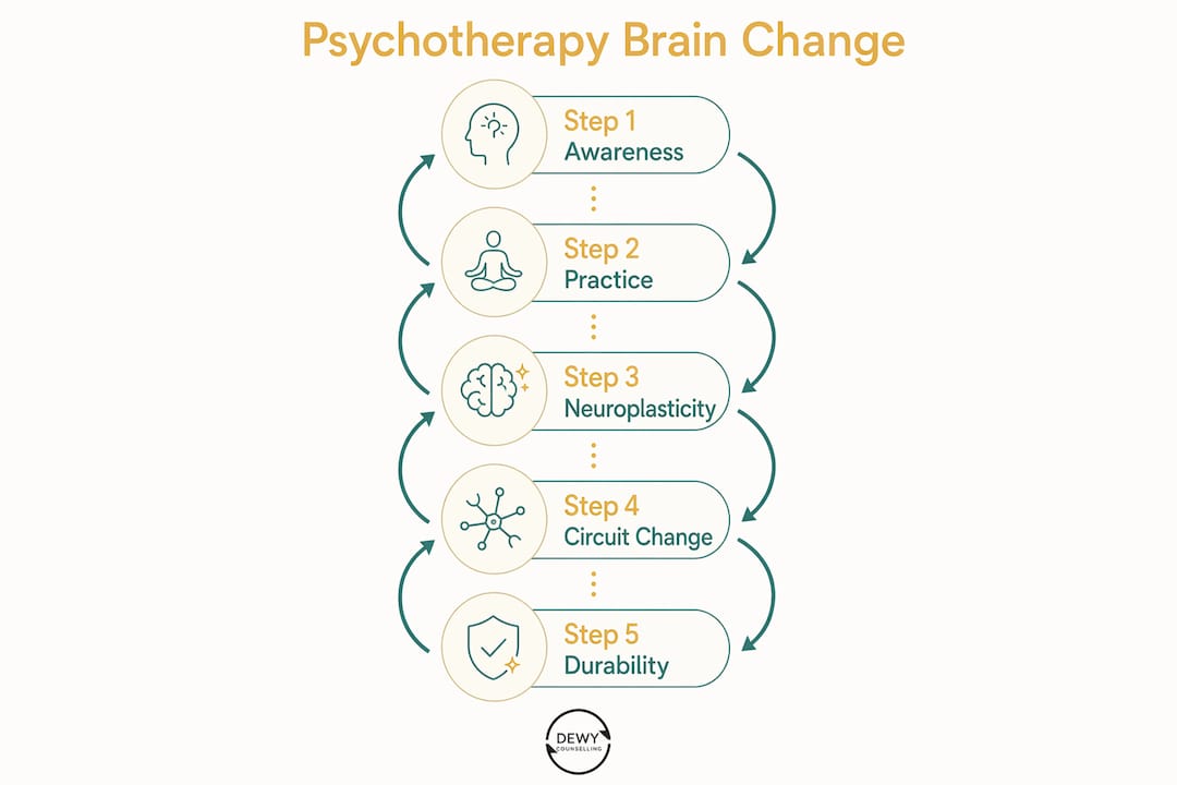
What meta-analyses show: Across depression and anxiety disorders, post-therapy neuroimaging consistently reveals decreased activation in the ACC, insula, and inferior frontal gyrus, regions associated with hyperarousal, rumination, and threat bias. These changes partially normalize the neural patterns seen in untreated illness, providing an objective biological account of symptom improvement.
| Study type | Key finding | Typical timepoint |
|---|---|---|
| Meta-analyses (depression/anxiety fMRI) | Decreased ACC, insula, IFG activation post-therapy | End of treatment |
| Longitudinal task-based fMRI (CBT) | Increased PFC engagement; reduced limbic reactivity | weeks to months |
| RCT with neuroimaging substudy (computer-augmented CBT) | Increased PFC-subcortical connectivity; symptom reduction | weeks to months |
| Resting-state fMRI (mindfulness/MBCT) | Altered DMN connectivity; reduced rumination-related activity | weeks to months |
| Long-term follow-up (pooled analyses) | Durable relapse reduction vs. medication alone | months post-treatment |
Timeline for change: Longitudinal imaging studies report that early functional changes, shifts in task-related activation patterns, can appear within weeks to months of starting therapy. Structural and connectivity changes tend to require sustained practice and longer follow-up to become reliably measurable. This mirrors what clinicians observe: mood and anxiety symptoms often improve before the deeper behavioral and relational patterns shift.
Durability: Pooled follow-up analyses show a relative risk of relapse of approximately 0.58 for psychotherapy compared to pharmacotherapy alone in major depressive disorder, meaning therapy roughly halves the risk of relapse compared with medication alone. This durability advantage likely reflects the fact that therapy builds new cognitive and behavioral skills that persist after treatment ends, whereas medication effects depend on continued use.
How psychotherapy compares with medication for brain change
Therapy and medication both work, and they often work on overlapping circuits, but they do not work identically. Understanding the distinction helps you and your clinician make a more informed choice.
- Overlapping but not identical effects: Both antidepressants and CBT reduce amygdala hyperreactivity and improve prefrontal regulation, but they approach this from different directions. Medication modulates neurotransmitter availability (serotonin, norepinephrine, dopamine), which shifts circuit function chemically. Therapy drives the same circuits through learning and experience, building new pathways rather than chemically adjusting existing ones.
- Durability advantage for therapy: The pooled relapse data consistently favor psychotherapy for long-term prevention, particularly in depression. Skills learned in therapy remain accessible after treatment ends; medication effects typically taper when the prescription stops.
- When combination treatment helps: For moderate-to-severe depression, bipolar disorder, OCD, and psychosis-spectrum conditions, combining therapy with medication often produces additive benefits. Medication can reduce symptom severity enough to make the cognitive and emotional work of therapy more accessible, while therapy builds the skills that sustain gains after medication is eventually tapered.
- When therapy alone is appropriate: Mild-to-moderate depression and anxiety, specific phobias, PTSD, and many adjustment disorders respond well to therapy alone, particularly when the patient prefers to avoid medication or has had adverse reactions previously.
- When medication alone may be prioritized: Severe acute episodes, conditions with strong biological loading (e.g., bipolar I, schizophrenia), or situations where the patient cannot engage meaningfully in therapy due to symptom severity may warrant medication as the primary initial intervention.
For readers interested in how these choices play out specifically for depression, psychotherapy for depression is worth reading alongside this article.
Pro Tip: If you are weighing therapy versus medication, ask your clinician specifically about relapse rates for your diagnosis, not just acute response rates. The durability data often favor therapy or combination treatment in ways that short-term symptom scores do not capture.
Mental health treatment in recovery contexts often requires this same integrated thinking, and integrated dual diagnosis treatment illustrates how combined approaches are applied in practice.
How researchers study brain change and what the limits are
The neuroimaging evidence is compelling, but reading it well means understanding how it is produced and where it falls short.
Study designs: Most research uses pre-and-post longitudinal imaging, comparing brain activity or connectivity before and after a course of therapy. Task-based fMRI measures brain responses to specific stimuli (emotional faces, threat cues, cognitive tasks). Resting-state fMRI captures spontaneous network activity without a task. PET receptor studies can track neurotransmitter system changes. RCT imaging substudies add a control group, strengthening causal inference.
Key limitations: Small sample sizes are pervasive in neuroimaging research, partly because scanning is expensive and logistically demanding. Heterogeneity across studies, in terms of patient populations, therapy protocols, imaging tasks, and outcome measures, makes direct comparison difficult. Most studies mix treatment responders and non-responders in their analyses, which can obscure the neural signature of genuine change. Publication bias toward positive findings is a real concern.
"Neuroimaging can reliably tell us about group-level circuit changes associated with successful therapy. What it cannot yet do is tell us, for any individual patient, which treatment will work best based on their brain scan alone. That predictive capacity remains a research goal, not a clinical tool."
— The effects of psychotherapy on brain function: A systematic and critical review
Causal inference is also genuinely difficult. Showing that brain activity changes after therapy does not prove that the therapy caused the change, rather than natural recovery, placebo effects, or regression to the mean. The strongest evidence comes from RCTs with active control conditions and neuroimaging substudies, which remain relatively rare. Individual differences in genetics, age, baseline brain function, and prior treatment history further complicate interpretation.
What actually happens in sessions that drives brain change
The neuroscience becomes most useful when it translates into what you and your therapist actually do together. The mechanisms described above are not abstract; they are activated by specific session-level practices.
- Behavioral activation: Scheduling and completing meaningful activities counters the withdrawal and anhedonia of depression by engaging reward circuitry in the striatum and rebuilding motivational pathways.
- Cognitive restructuring: Identifying and challenging distorted thoughts strengthens PFC engagement and gradually weakens the automatic, fear-driven appraisals generated by the amygdala and insula.
- Guided exposure: Systematically approaching feared situations or memories activates the fear circuit in a controlled context, allowing extinction learning to update the amygdala's threat predictions.
- Trauma processing (EMDR, trauma-focused CBT): Reprocessing traumatic memories in a safe relational context facilitates reconsolidation, updating the emotional valence and contextual framing of stored memories through hippocampal and prefrontal mechanisms.
- Mindfulness practice: Sustained attention to present-moment experience, including bodily sensations and thoughts without judgment, modulates DMN activity and builds insula-based interoceptive awareness.
- Interpersonal and relational work: The therapeutic alliance itself is a neurobiological event. Feeling genuinely understood by another person activates social reward circuits and can modulate the stress-response systems that underlie many anxiety and trauma presentations.
Repetition is the key variable across all of these. A single session of cognitive restructuring does not remodel a circuit. Consistent practice, both within sessions and through between-session homework, is what consolidates new pathways. This is why weekly therapy sessions tend to produce better outcomes than sporadic contact: the brain needs repeated activation of the new pathway to strengthen it.
Pro Tip: Three evidence-based ways to support brain change between sessions: (1) practice the specific skill your therapist assigned, even briefly, every day; (2) prioritize sleep, since memory consolidation and synaptic pruning occur primarily during slow-wave sleep; (3) add moderate aerobic exercise, which promotes hippocampal neurogenesis and supports the mood regulation gains made in therapy.
How therapy improves daily life offers a practical companion to these session-level strategies for readers who want concrete examples.
Key Takeaways
Psychotherapy produces measurable, durable neuroplastic changes in emotion-regulation circuits, with the strongest evidence pointing to decreased limbic reactivity and increased prefrontal control across multiple disorders and therapy types.
| Point | Details |
|---|---|
| Therapy is a biological intervention | Psychotherapy drives experience-dependent synaptic change, altering functional connectivity in PFC-limbic circuits. |
| Multiple regions are affected | PFC, amygdala, ACC, insula, hippocampus, and striatum all show measurable changes after successful therapy. |
| Change takes weeks to months | Early functional shifts appear within weeks; structural and connectivity changes require sustained practice over months. |
| Therapy reduces relapse risk | Pooled follow-up data show a substantial reduction in relapse risk for psychotherapy compared to medication alone. |
| Dewycounselling applies these principles | Individual, couples, and family therapy at Dewycounselling is grounded in the same neuroscience-informed practices described here. |
The science matters, but so does what happens between two people
The neuroimaging literature has done something genuinely important: it has given us an objective account of why therapy works that goes beyond self-report. When a patient says they feel less afraid, less stuck, or more like themselves after a course of treatment, we now have brain-scan evidence that something real has shifted in the circuits underlying those experiences. That is meaningful, both for reducing stigma and for motivating people to stay with the process when it feels slow.
What the science cannot fully capture, though, is the role of the therapeutic relationship itself. The alliance between therapist and patient is not just a delivery mechanism for techniques. It is, in its own right, a neurobiological experience: one that activates social reward circuits, modulates the stress-response system, and creates the felt safety that makes genuine emotional processing possible. No fMRI study has fully quantified what it means to feel truly heard by another person, but clinically, that experience is often what makes the difference between a patient who engages with the hard work of therapy and one who drops out after three sessions.
The other thing worth saying plainly: brain change from therapy is gradual and individualized. Genetics, age, baseline brain function, prior trauma history, and the specific disorder all shape how quickly and how completely the brain responds. Realistic hope is not the same as guaranteed rapid transformation. Progress in therapy often looks like two steps forward and one step back, and that is consistent with how neuroplasticity actually works. Circuits do not remodel in a straight line. They strengthen through repeated practice, setback, and renewed effort, which is exactly what good therapy holds space for.
Ready to experience what therapy can do for your brain?
The science described in this article is not just academic. It is the foundation of what happens in a well-conducted therapy session, whether you are working through anxiety, depression, relationship patterns, or the aftermath of trauma. Dewycounselling offers individual, couples, and family therapy grounded in evidence-based, neuroscience-informed approaches, available both online and in person to fit your life.

Getting started is straightforward. You can reach out through Dewycounselling's services page to learn about available formats and book an initial session. If you are specifically interested in how therapy can support a relationship, couples counselling is also available. The first step is simply making contact. From there, the work, and the brain change it drives, can begin.
Visit Dewycounselling to learn more or schedule your first session.
Useful sources and further reading
These are the primary studies and reviews that underpin the science in this article. The first two are the most accessible starting points for lay readers; the remainder are best suited to clinicians and researchers.
- Meta-analyses of the neural mechanisms and predictors of response to psychotherapy in depression and anxiety — The most comprehensive meta-analytic synthesis of neuroimaging findings across therapy types; the best single source for understanding region-level changes and the dual-process model.
- Rebuilding the brain with psychotherapy — Grounds the field in Kandel's theoretical framework; accessible to educated non-specialists and essential for understanding why therapy qualifies as a biological intervention.
- The effects of psychotherapy on brain function: A systematic and critical review — Covers transdiagnostic effects and methodological limitations; recommended for clinicians wanting a balanced appraisal of what neuroimaging can and cannot tell us.
- Brain functional effects of CBT for depression: A systematic review of task-based fMRI studies — Focused specifically on CBT and depression; the clearest source for timeline data on when functional changes become detectable.
- Neuroimaging changes in major depression with brief computer-assisted CBT compared to waitlist — RCT evidence for PFC-subcortical connectivity changes with a brief, technology-assisted CBT protocol; relevant for clinicians and patients interested in shorter-format interventions.
- Psychotherapy offers durable long-term benefits often exceeding pharmacotherapy alone — The primary source for relapse and recurrence data comparing therapy with medication; essential reading for anyone weighing treatment options.
- Neural correlates of psychodynamic and non-psychodynamic therapies: A meta-analysis and systematic review — Covers fMRI findings across therapy modalities including psychodynamic approaches; useful for clinicians working outside the CBT tradition.
This article is general educational information, not professional medical or psychological advice. Please consult a qualified clinician for guidance specific to your situation.